Export
Record an 8-second clip: the waveform alone, waveform plus spectrogram, or the whole screen, with sound when audio is on.
Record an 8-second clip: the waveform alone, waveform plus spectrogram, or the whole screen, with sound when audio is on.
A 0–100 depth surrogate from the model's own spectral edge and suppression ratio, smoothed over fifteen seconds so it lags the trace as a device's number does.
A live, pixel-identical copy of the monitor on a projector or second screen, or streamed to up to 30 learner laptops that join with a session code, while you drive the case.
Adjust gain, sweep speed, the spectrogram's colour window and the spectral-edge percentile to see how a setting changes what you read. Add movement, ECG pulse, 50 Hz mains or diathermy artifact, with the unit's activation tone when audio is on, and see what each does to the trace, the spectrogram and the numbers. Reset to device calibration in one click.
Adult ASA I to IV, about 45 to 90 years, on one axis. Amplitudes fall, alpha thins, rhythms slow, and the same dose reads deeper, down to burst suppression.
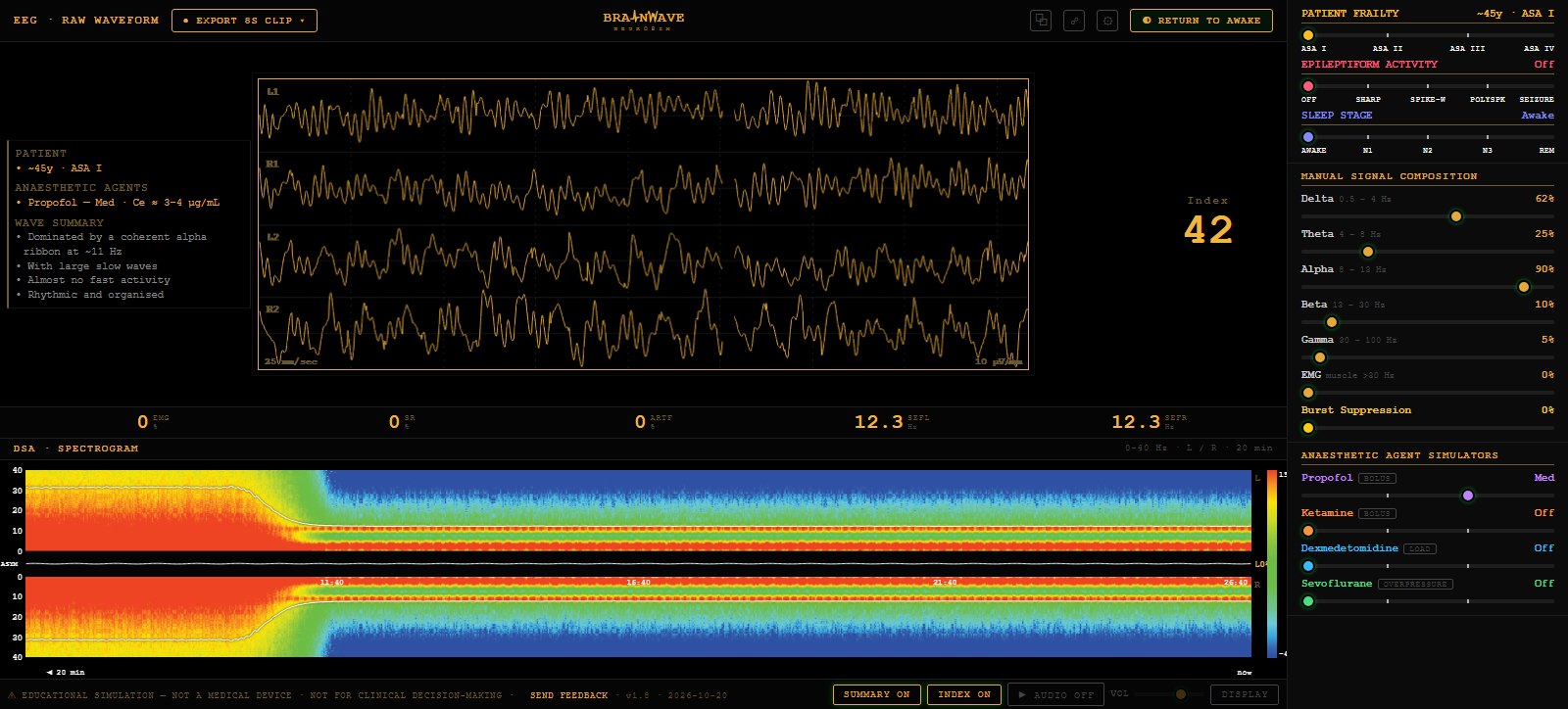
L1, R1, L2 and R2 in real time on the default display (a single channel on Compact), calibrated in µV/mm and mm/sec. Blinks, movement and every drug and patient effect show in the morphology.
A plain-language description of what the trace shows, typed in once the settings settle. Hover a line for its teaching note and watch the matching wave light up on the trace.
EMG, suppression ratio, artifact and the spectral edge for each hemisphere, each with a hover teaching note.
Sharp waves, 3 Hz spike-wave, polyspike and an evolving seizure, each followed by postictal slowing and each with its own spectrogram flame.
N1, N2, N3 and REM: spindles, K-complexes, sawtooth waves, slow and rapid eye movements, with the drugs locked out.
Drive delta, theta, alpha, beta, gamma, EMG and burst suppression directly, to build or isolate any pattern.
0–40 Hz, left over right, a 20-minute window at twenty times real time so a whole induction fits one screen. Spectral-edge trace, asymmetry graph, suppression and artifact columns, injection ticks.
Hear the trace. Each band pulses at its own rate, burst suppression gates the sound and EMG hisses over it.
Switch between the Standard four-channel layout and the Compact index-led layout, with a single-panel spectrogram, an index trend and a number column.
Propofol, ketamine, dexmedetomidine and sevoflurane at Off, Low, Med and High, blended through a response-surface interaction model. Bolus, loading-dose and overpressure buttons give the transient. Return to Awake resets the patient.
Every signal is computer-generated and modelled on adult patients. Not a medical device and not for any patient-care decision.
Try the demo, or read the why and what it’s based on.