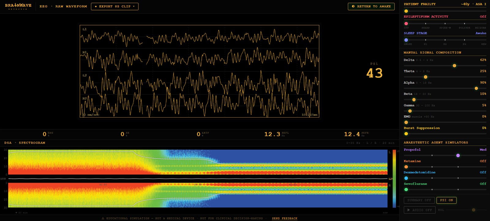
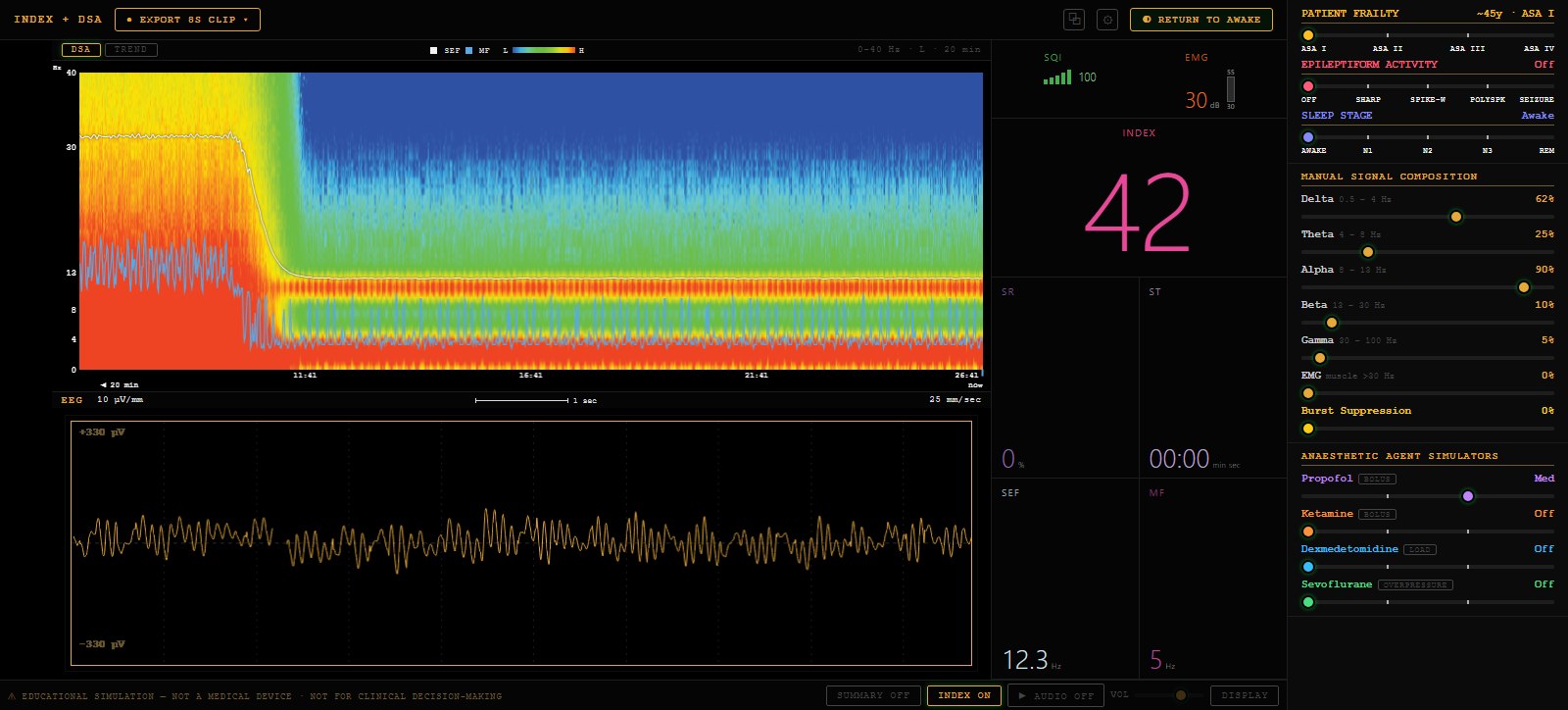
Export
8-second clip: waveform, EEG + DSA, or full window.
Mirror window & laptop link
Duplicate the monitor on a projector, or stream it live to up to 30 learner laptops with a session code.
Summary panel
Plain-language description of the live trace.
Parameters
EMG · SR · ARTF · SEFL / SEFR.
DSA spectrogram
0–40 Hz, L over R, 20-minute window.
Index — depth readout
0–100, derived from the raw EEG, spectral edge and suppression ratio.
Patient frailty
Age and comorbidity on one axis.
Epileptiform activity
Sharp · spike-wave · polyspike · seizure, with postictal slowing.
Sleep stage
N1 · N2 · N3 · REM, with spindles, K-complexes, sawtooth and eye movements.
Manual composition
Drive δ θ α β γ, EMG and burst suppression directly.
Anaesthetic agents
Propofol · ketamine · dexmedetomidine · sevoflurane, with a response-surface interaction model.
Multiple display types
Standard four-channel, or Compact index-led with a number column and index trend.
Audio
Sonify the displayed waveform.